Kidney Cancer
From Kidney Cancer Resource
Overview
Renal cell carcinoma (RCC, aka hypernephroma) is the most common form of kidney cancer arising from the proximal renal tubule. It is the most common type of kidney cancer in adults. Initial treatment is most commonly a radical or partial nephrectomy. It is generally resistant to radiation therapy and chemotherapy, although some cases respond to immunotherapy.
To read one of the most useful & knowledgeable accounts of Kidney Cancer Staging and grading, see Steve Dunn's site, himself a kidney cancer stage 4 survivor for 15 years, Steve has been an inspiration to countless thousands of Kidney Cancer Patients around the world. His death in 2005 from bacterial meningitis was a tragedy. Steve's words are quoted in various places on this site.
Kidney Cancer Videos
We offer a choice of links to view Kidney Cancer Videos .
Normal Anatomy
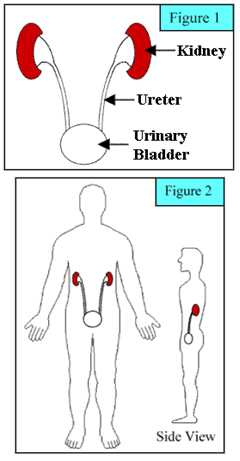
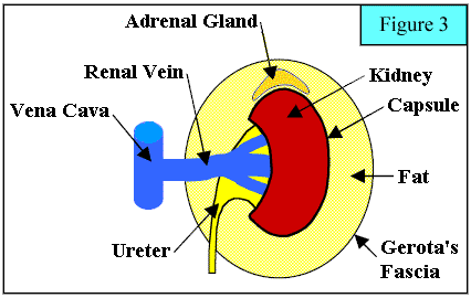
Most people have two Kidneys. The Kidneys produce urine, which drains through narrow tubes (called Ureters) into the urinary bladder (Figure 1). The Kidneys are located toward the back of the flank, with one Kidney on either side (Figure 2). The Kidney is contained within a fibrous sheath called Gerota’s Fascia (Figure 3). Within this fascia is a layer of fat that surrounds the Kidney. The capsule is a thin layer that covers the outer surface of the Kidney (analogous to the red external layer of an apple). The primary vein that drains the kidney (renal vein) merges with the vein that takes blood to the heart (Vena Cava). The term “renal” means pertaining to the Kidney. An Adrenal gland is located above each Kidney within Gerota's Fascia.
Several types of tumours both benign and malignant may occur in the Kidney. A Kidney Tumour is an abnormal area within the Kidney. The terms mass, lesion, and tumor are often used interchangeably. Tumours may be Benign (not cancerous) or Malignant (cancerous). The most common type of Kidney Tumour is a fluid-filled area called a Cyst(see Bosniak Rating). Simple Cysts are Benign and have a typical appearance on imaging studies. Simple cysts do not progress to cancer and usually require no follow-up treatment. Complex Cysts do not have the typical benign appearance and may contain cancer. When Complex Cysts are present, the need for treatment is determined on an individual basis. Another type of Kidney Tumour is a solid Kidney Tumour (i.e. not fluid-filled). Solid Kidney Tumours may be Benign, but are usually Malignant. In fact, more than 90% of solid Kidney Tumours are cancerous.
In the United States, Kidney Cancer accounts for about 3% of all cancers, with approximately 12,000 Kidney Cancer deaths each year. In Britain currently around 6,600 Diagnosese of Kidney Cancer are recorded annually and there is an admission that some 3,600 per annum lead to death, this latter figure is however felt to be deeply suspect and is thought to be much higher, though masked by subsequent problems where the roots are in fact in the original disease. Kidney Cancer occurs slightly more often in males and is usually diagnosed between the ages of 50 and 70, but can occur at any age. In adults, the most common type of kidney cancer is Renal Cell Carcinoma, also called renal adenocarcinoma or hypernephroma.
Kidney Cancer Types
KC Symptoms
For more details on Symptoms Click Here
KC Causes
For more details of Causes Click Here
Detecting Kidney Cancer
When a Kidney Tumour is suspected, a Kidney imaging study is obtained. The initial imaging study is usually an Ultrasound, CT Scan or MRI Scan. In some cases, a combination of imaging studies may be required to completely evaluate the Tumour. If cancer is suspected, the patient should be evaluated to see if the cancer has spread beyond the Kidney. Renal Cell Carcinoma is sometimes diagnosed with a Biopsy.
Generally Biopsy of the kidney is discouraged for a number of reasons, including possible false negative readings, the vascularity of the tissue and the fact that if cancerous it can, in very rare cases spread along the needle's exit path.
During a Biopsy, a physician removes a sample of tissue for examination under the microscope. When the cancer is examined under the microscope, there are two distinct cell types: Clear Cell and Granular Cell (Sarcomatoid), with some cancers being a mixture of both cell types. Cell types generally do not influence outcome following treatment; however, in some clinical studies, treatment with Proleukin® results in better outcomes for patients with Clear Cell Carcinoma.
KC Staging
For details on the Staging of Kidney Cancer goto this link
KC Grading
For details on the Grading of Kidney Cancer goto this link
KC Treatment
For more details on Treatment Click Here
KC Drugs
Please click here for more detail on the available drugs for Kidney Cancer
KC Prognosis
For more details on Prognosis Click Here
KC Follow-Up
For more details on Treatment Click Here
Kidney Cancer Advances
Kidney Cancer Association's Clinical Highlights from The Fifth International Kidney Cancer Symposium Click Here
Kidney Cancer MD Anderson (Oncology)
See Also
References
|
Disclaimer
Kidney Cancer Resource (KCR) is not influenced by sponsors. The information contained herein is not intended as a substitute for the advice of an appropriately qualified and licensed physician or other licensed health care provider. The information provided here is for educational and information purposes only. Early accurate Diagnosis (Dx.) saves lives. Please check with a physician if you suspect you are ill, never ignore Symptoms. To help your health care specialist make an accurate Diagnosis please keep notes of dates, times and details of your Symptoms. We are not offering medical advice nor do we consider links, individuals or articles accessed through this site to be offering medical advice.
E&OE - Errors & Omissions Excepted
As much of the information posted on this Web Site for peoples convenience is of a medical or technical nature, and may be a matter of life or death the E&OE is a Disclaimer showing that to the best of our ability information is accurate and correctly written or transcribed. Before acting on information on this site you are responsible for checking it with your relevant medical team. We can not be held responsible for any Errors & Omissions made; nor for information on links and articles provided in good faith.


